Glaucoma at a Glance
Glaucoma is a group of eye diseases that damage the optic nerve, typically driven by elevated intraocular pressure (IOP), and it is the leading cause of irreversible blindness worldwide. Most early-stage cases cause no pain and no noticeable vision change; damage accumulates silently for years before peripheral vision loss becomes apparent. The one exception is acute angle-closure glaucoma, which is a medical emergency: sudden severe eye pain, halos around lights, and nausea require immediate emergency care, not a scheduled appointment. Early detection through a comprehensive dilated eye exam remains the only reliable way to catch glaucoma before significant vision is lost.
This article is for informational purposes only and is not a substitute for a comprehensive ophthalmologic evaluation. If you have concerns about your eye pressure or vision, see an ophthalmologist.
Why Glaucoma Is Called the Silent Thief of Sight
Glaucoma qualifies for that description because primary open-angle glaucoma (POAG, the most common form) progresses without warning signs. According to the National Eye Institute (NEI), open-angle glaucoma is painless and causes no vision changes at first. Blind spots develop slowly in peripheral (side) vision, and most people do not notice any change until the damage is quite severe.
This pattern has serious public health consequences. According to a systematic review and meta-analysis published in Ophthalmology (Tham et al., 2014), approximately 76 million people globally had glaucoma in 2020, with that number projected to reach 111.8 million by 2040. Glaucoma is the most common cause of irreversible blindness worldwide. A large proportion of affected individuals remain undiagnosed because the disease gives no early signal.
The distinction between “no symptoms now” and “no damage now” matters clinically. By the time a patient notices peripheral field loss, they have often already lost 25 to 40% of their retinal ganglion cells.
Types of Glaucoma: What Clinicians and Patients Need to Know
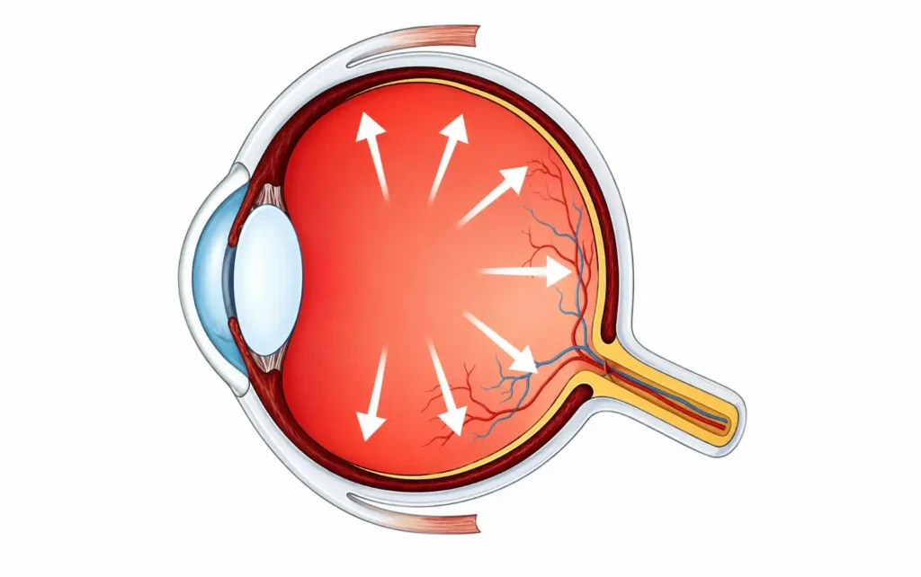
Glaucoma is not a single disease. The American Academy of Ophthalmology (AAO) describes glaucoma as a disease that damages the eye’s optic nerve, usually when fluid builds up in the front part of the eye, raising intraocular pressure and damaging the optic nerve over time.
The major types differ in mechanism, presentation, and urgency:
Primary open-angle glaucoma (POAG, the most common slow-progressing form) is the most common form worldwide. The drainage angle between the iris and cornea remains open, but the trabecular meshwork (the eye’s internal drainage filter) gradually becomes less efficient. IOP rises slowly, and optic nerve damage accumulates without symptoms. POAG accounts for the majority of glaucoma cases in populations of European and African descent.
Primary angle-closure glaucoma (PACG) occurs when the iris physically blocks the drainage angle. It is disproportionately common in people of East and Southeast Asian ancestry, as well as those with hyperopia (farsightedness). PACG can be chronic and relatively silent, or it can present as an acute attack, which is a true ophthalmic emergency.
Normal-tension glaucoma (NTG, optic nerve damage despite normal-range eye pressure) is a subtype of open-angle glaucoma in which optic nerve damage occurs despite IOP that remains within the statistically normal range (typically 10-20 mmHg). According to the NEI, scientists are not certain why this happens, though vascular factors and optic nerve vulnerability are suspected. NTG is considerably more common among Japanese populations.
Secondary glaucoma results from another condition: uveitis, trauma, prolonged corticosteroid use, diabetic neovascularization (neovascular glaucoma), or pigment dispersion syndrome (pigmentary glaucoma). Identifying and treating the underlying cause is part of the management.
Congenital glaucoma presents in infants and young children, usually from a developmental abnormality in the drainage angle. It requires surgical intervention.
Comparing Primary Open-Angle vs. Primary Angle-Closure Glaucoma
The clinical distinction between POAG and PACG matters for triage and treatment planning.
| Feature | Primary Open-Angle Glaucoma | Primary Angle-Closure Glaucoma |
|---|---|---|
| Drainage angle | Open | Closed or narrowed |
| Onset | Gradual, often over years | Chronic (gradual) or acute (sudden) |
| Symptoms | Usually none until late-stage peripheral loss | Acute: severe eye pain, headache, halos, nausea, blurred vision |
| IOP | Elevated or normal (NTG) | Often markedly elevated during acute attack |
| Urgency | Routine specialist referral; monitor progression | Acute attack: emergency room immediately |
| Common populations | All; higher prevalence in Black/African American patients | East/Southeast Asian ancestry; hyperopes |
| Primary treatment | IOP-lowering drops, SLT laser, surgery | Laser peripheral iridotomy; acute IOP crisis management |
Risk Factors: Who Should Be Screened Earlier
Glaucoma risk is not evenly distributed. The NEI identifies these established risk factors for primary open-angle glaucoma:
- Age over 60: Risk rises significantly after 60 for the general population, and after 40 for African Americans.
- Race and ethnicity: Black/African American individuals have approximately a threefold higher prevalence of open-angle glaucoma compared to non-Hispanic whites, are diagnosed at younger ages (mean around 60 vs. 66 years), and experience more rapid progression, according to research published in the American Journal of Ophthalmology (Keel et al.). Hispanic/Latino patients also face earlier onset and higher severity. East and Southeast Asian populations carry elevated risk specifically for angle-closure glaucoma.
- Family history: A first-degree relative with glaucoma substantially increases individual risk.
- High myopia: Axial length elongation in highly myopic eyes is associated with elevated glaucoma risk. Refractive errors like high myopia change the structural relationship between the optic disc and surrounding tissue. For patients with myopia and astigmatism together, the higher the sphere, the more important annual retinal evaluation becomes.
- Diabetes mellitus: Diabetic patients have elevated glaucoma risk, likely through vascular and structural pathways.
- Long-term corticosteroid use: Topical, systemic, and inhaled corticosteroids can elevate IOP in susceptible individuals (“steroid responders”).
- Prior eye trauma: Angle recession from blunt trauma can impair drainage and cause secondary glaucoma years after the original injury.
- Elevated IOP (ocular hypertension): IOP above 21 mmHg without structural optic nerve damage or visual field loss is termed ocular hypertension. It is a risk factor but not the same as glaucoma: not everyone with elevated IOP develops glaucoma, and glaucoma can occur at normal pressures (as in normal-tension glaucoma). IOP is one piece of the diagnostic picture, not the sole criterion.
Glaucoma Symptoms by Type
Open-Angle Glaucoma: No Reliable Early Warning
Most open-angle glaucoma is found during a routine dilated eye exam, not because the patient noticed anything wrong. The earliest losses occur in the peripheral visual field, areas that the brain partially compensates for using the opposite eye. By the time a patient spontaneously notices peripheral field loss, the disease is typically in a moderate to advanced stage.
This is why visual acuity testing alone does not detect early glaucoma. Central acuity can remain 20/20 while significant peripheral and paracentral field damage accumulates.
Angle-Closure Glaucoma: Recognize the Emergency
Acute primary angle-closure presents with a characteristic cluster of symptoms. As described in StatPearls (NCBI Bookshelf):
Acute angle-closure glaucoma presents as a sudden onset of severe unilateral eye pain or headache associated with blurred vision, rainbow-colored halos around bright lights, nausea, and vomiting.
The pupillary response is also affected: the pupil may be mid-dilated and unreactive during an acute attack, and light sensitivity can be pronounced.
Emergency Triage: Know the Difference
Go to the emergency room immediately if you or a patient experiences sudden severe eye pain, headache, halos around lights from one eye, nausea or vomiting, and blurred vision together. This presentation is consistent with acute angle-closure glaucoma, which can cause permanent vision loss within hours without treatment.
Schedule an urgent ophthalmologist appointment (within days to a few weeks) if you notice gradual loss of side (peripheral) vision, particularly if you are in a high-risk group. Gradual peripheral loss is the typical presentation of open-angle glaucoma and warrants prompt evaluation, but it is not the same emergency.
How Glaucoma Is Diagnosed
Glaucoma diagnosis requires a comprehensive eye examination. No single test is sufficient. An ophthalmologist will typically perform these steps in combination:
- **Tonometry** measures intraocular pressure, most accurately using applanation tonometry. An IOP reading above 21 mmHg raises concern, but normal IOP does not rule out glaucoma. The Baltimore Eye Survey (Sommer et al., 1991) found that approximately half of patients with primary open-angle glaucoma had IOP readings below 22 mmHg at their screening visit. The Ocular Hypertension Treatment Study (Kass MA et al., [Arch Ophthalmol 2002](https://www.aoa.org/assets/documents/EBO/931-094KASS-2002.pdf)) found that only 9.5% of patients with elevated IOP who were left untreated for five years developed early glaucoma, underscoring that elevated IOP alone is not the same as glaucoma.
- **Ophthalmoscopy / fundus examination** evaluates the optic disc. Key findings include enlargement of the optic nerve cup relative to the disc (cup-to-disc ratio greater than 0.5 is a flag), focal or diffuse neuroretinal rim thinning (especially in the superior or inferior quadrant), and disc hemorrhages.
- **Perimetry** (visual field testing — a test where you press a button each time you see a flashing light in your peripheral vision) detects functional damage. Automated white-on-white perimetry is the gold standard for detecting and tracking visual field defects. Establishing a baseline is essential, since glaucoma management depends on tracking progression over time.
- **Optical coherence tomography (OCT)** provides high-resolution structural imaging of the optic nerve and retinal nerve fiber layer (RNFL — the thin layer of nerve fibers that transmit visual signals from the retina to the brain). OCT can detect structural changes before they produce measurable visual field loss, making it especially valuable for early diagnosis and monitoring.
- **Gonioscopy** (an in-office exam of the eye’s drainage angle using a contact-lens mirror) examines the anterior chamber angle directly. It is the gold standard for distinguishing open-angle from angle-closure glaucoma and is essential before laser or surgical intervention.
Glaucoma can also cause acquired color vision deficiency, particularly affecting blue-yellow discrimination. A color vision test as part of the baseline assessment can help track functional changes over time.
Glaucoma Treatment: A Tiered Approach
The goal of treatment is to preserve vision by lowering IOP to a level at which optic nerve damage stops progressing. Treatment does not restore lost vision; it prevents further loss. The AAO’s preferred practice patterns provide the clinical framework for POAG management.
Medications (First-Line for Most Patients)
Prostaglandin analogs (latanoprost, bimatoprost, travoprost) are the most commonly prescribed first-line agents. They lower IOP by increasing aqueous humor outflow through the uveoscleral pathway (an alternate drainage route that bypasses the trabecular meshwork). Once-daily dosing and strong efficacy make them the preferred starting point; clinical trials consistently show IOP reductions of 25 to 33% from baseline.
Beta-blockers (timolol) reduce aqueous production and are effective, delivering roughly 20 to 25% IOP reduction as monotherapy, though they carry systemic risks in patients with asthma, bradycardia, or heart block.
Alpha-2 agonists (brimonidine) reduce aqueous production and increase uveoscleral outflow. They may have a neuroprotective role, though evidence on that point is still developing.
Carbonic anhydrase inhibitors (dorzolamide topically, acetazolamide systemically) reduce aqueous production by about 15 to 20%. Systemic carbonic anhydrase inhibitors are typically reserved for acute situations due to side effects.
Rho kinase inhibitors (netarsudil) are a newer class that increase trabecular outflow and reduce episcleral venous pressure.
Laser Treatment
Selective laser trabeculoplasty (SLT) applies low-energy laser pulses to the trabecular meshwork to improve drainage. The landmark LiGHT trial, a multicenter randomized controlled trial published in The Lancet (2019), found that 74.2% of patients treated with first-line SLT were drop-free at 36 months, with 95% achieving target IOP. SLT is now widely considered a valid first-line alternative to medication for newly diagnosed open-angle glaucoma and ocular hypertension.
Laser peripheral iridotomy (LPI) is the primary treatment for angle-closure glaucoma. A laser creates a small opening in the peripheral iris, allowing aqueous humor to bypass the blocked drainage angle and restore normal flow. It is performed urgently in acute angle closure and prophylactically in eyes with narrow angles at risk.
Surgery
Trabeculectomy creates a controlled drainage pathway (a small flap in the sclera) that bypasses the trabecular meshwork. It remains the most established surgical option when medications and laser fail to adequately control IOP.
Tube shunt surgery (glaucoma drainage devices) implants a small tube that drains aqueous to an external reservoir plate. Used when trabeculectomy has failed or is likely to fail.
Minimally invasive glaucoma surgery (MIGS) encompasses a growing family of procedures (iStent, Hydrus microstent, gonioscopy-assisted transluminal trabeculotomy) that aim to lower IOP with a better safety profile than traditional surgery. MIGS procedures are typically combined with cataract surgery and are most appropriate for mild to moderate glaucoma.
Glaucoma Screening: How Often and for Whom
The NEI states that a comprehensive dilated eye exam is the only reliable method for early glaucoma detection. The following frequency recommendations are drawn from NEI and AAO guidance:
| Population | Recommended Screening Frequency |
|---|---|
| General adults 40-54, no risk factors | Every 2-4 years |
| General adults 55-64, no risk factors | Every 1-3 years |
| Adults 65 and older | Every 1-2 years |
| African Americans age 40 and older | At least every 2 years |
| Adults with family history of glaucoma | At least every 2 years from age 40 |
| Adults with diabetes or hypertension | As directed by physician, at least every 2 years |
| High myopes (over -6.00 D) | Annual evaluation recommended |
| Anyone with prior ocular hypertension | As directed by ophthalmologist, typically annually |
Prognosis: What Early Detection Actually Changes
Glaucoma-related vision loss is irreversible. The retinal ganglion cells that form the optic nerve do not regenerate. This is what makes early detection so consequential: treatment initiated before significant field loss can slow or halt progression indefinitely in most patients.
The NEI states directly that there is no cure for glaucoma, but early treatment can often stop the damage and protect vision. The goal is not recovery; it is preservation.
For patients diagnosed in early stages, current medical and surgical tools make it entirely possible to maintain functional vision for life. For patients diagnosed after significant loss, the treatment goals shift to slowing further damage and optimizing remaining function.
Frequently Asked Questions
Can glaucoma be cured?
No. Glaucoma cannot be cured. According to the National Eye Institute, there is no cure for glaucoma, but early treatment can often stop the damage and protect vision. Existing damage to the optic nerve is permanent, as retinal ganglion cells do not regenerate. Treatment (whether medications, laser, or surgery) controls IOP to prevent further loss but does not restore what has already been lost.
Can you go blind from glaucoma?
Yes, untreated or inadequately treated glaucoma can cause complete blindness. Glaucoma is the leading cause of irreversible blindness worldwide. However, in most cases, vision loss can be significantly slowed or halted with treatment. The risk of blindness is substantially lower for patients who are diagnosed early and maintain good IOP control with consistent follow-up.
Is glaucoma hereditary?
A family history of glaucoma is a recognized risk factor for developing the disease. First-degree relatives of people with glaucoma face a significantly elevated risk. However, glaucoma is not strictly inherited in a simple Mendelian pattern; it is a complex disease influenced by multiple genetic and environmental factors. Having a relative with glaucoma means you should begin regular screening at age 40 rather than waiting until 60.
What is the difference between glaucoma and ocular hypertension?
Ocular hypertension means your IOP measures above 21 mmHg without any optic nerve damage or visual field loss. Glaucoma means optic nerve damage is present, which may or may not be accompanied by elevated IOP. Some people with ocular hypertension never develop glaucoma; some people with normal IOP do develop glaucoma (normal-tension glaucoma). The distinction matters because not all ocular hypertension requires the same level of intervention.
At what age should I start screening for glaucoma?
The American Academy of Ophthalmology recommends a baseline comprehensive eye exam at age 40 for all adults. If you are African American, the National Eye Institute recommends beginning regular screening from age 40 due to significantly higher risk. If you have a family history of glaucoma, diabetes, high myopia, or a history of corticosteroid use, start discussing screening frequency with your eye care provider well before 40. Adults over 65 should have a comprehensive exam every one to two years regardless of risk factors.
Does glaucoma cause eye pain?
Primary open-angle glaucoma, the most common type, does not cause pain. This is one reason it is so often diagnosed late. Acute angle-closure glaucoma does cause severe, sudden eye pain, along with headache, halos around lights, nausea, and vomiting. If you experience those symptoms together, seek emergency care immediately. Pain alone is not a reliable indicator; most people with glaucoma feel nothing unusual until significant vision loss has occurred.
What eye drops are used for glaucoma?
The most commonly prescribed first-line drops are prostaglandin analogs (latanoprost, bimatoprost, travoprost), given once daily at night. Other classes include beta-blockers (timolol), alpha-2 agonists (brimonidine), carbonic anhydrase inhibitors (dorzolamide), and the newer rho kinase inhibitors (netarsudil). The choice depends on the degree of IOP reduction needed, the patient’s systemic health, and tolerability. Adherence to the prescribed schedule is critical: drops only work if taken consistently.
This article draws on clinical guidelines from the American Academy of Ophthalmology, the National Eye Institute, and peer-reviewed research indexed on PubMed. It is intended as an educational reference for patients and eye care professionals. It is not a substitute for examination, diagnosis, or treatment by a qualified ophthalmologist.

I am a seasoned software engineer with over two decades of experience and a deep-rooted background in the optical industry, thanks to a family business. Driven by a passion for developing impactful software solutions, I pride myself on being a dedicated problem solver who strives to transform challenges into opportunities for innovation.