
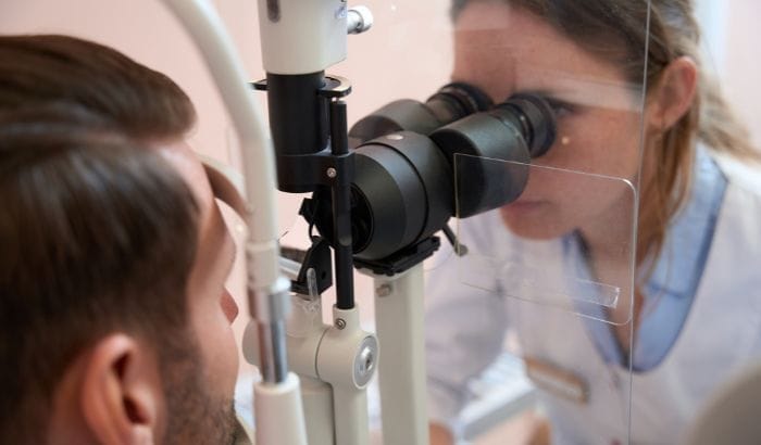
Quick Answer: Visual acuity is reported in four interchangeable notations: Snellen imperial (20/20), Snellen metric (6/6), decimal (1.0), and logMAR (0.0), which all describe the same standard level of vision. A result of 6/12 is the same as 20/40 or decimal 0.5, a mild reduction that still falls within the World Health Organization’s “no visual impairment” range. A result of 0.1 on the decimal scale is the same as 6/60 or 20/200, the U.S. legal blindness threshold. The table below shows how the four scales relate.
How to read visual acuity notation: Snellen, decimal, and logMAR
Visual acuity results show up in different formats depending on where and how the test was performed, and mistaking one notation for another is a common source of misread prescriptions and lab records. All four systems measure the same thing: how a patient’s smallest resolvable detail compares with a defined standard.
How the scales convert into each other
The Snellen fraction is a ratio of two distances. The top number is how far the patient stands from the chart (20 feet in the United States, 6 meters in most other countries), and the bottom number is how far a person with standard vision would need to stand to read that same line. Divide the two and the result is the decimal equivalent: 20/40 works out to 0.5, and 6/12 also works out to 0.5, because 20/40 and 6/12 describe the same line on the chart in different units.
LogMAR takes that decimal value one step further. It is the base-10 logarithm of its reciprocal: logMAR = log10(1 ÷ decimal). For a decimal of 0.5, that is log10(2), which rounds to 0.3. For a decimal of 0.1, log10(10) comes out to exactly 1.0. A 2010 study published in Arquivos Brasileiros de Oftalmologia (“Tables for measuring visual acuity on a logarithmic scale”) found that logMAR charts are the most appropriate scale for clinical follow-up and population-based visual acuity surveys, because the spacing between lines is uniform in a way the Snellen fraction is not.
Lower logMAR values mean better acuity, the opposite direction from decimal and unlike Snellen, where the numbers simply get further apart. Negative logMAR values, such as -0.1, indicate acuity above the standard reference.
Visual acuity conversion table
| Snellen (imperial) | Snellen (metric) | Decimal | logMAR | What it means |
|---|---|---|---|---|
| 20/20 | 6/6 | 1.0 | 0.0 | Standard reference vision |
| 20/40 | 6/12 | 0.5 | 0.3 | Mild reduction; still counts as “no visual impairment” under WHO criteria |
| 20/200 | 6/60 | 0.1 | 1.0 | U.S. legal blindness threshold |
What does 6/12 vision mean?
A 6/12 result (20/40 in the imperial system, 0.5 decimal, 0.3 logMAR) means the patient can read at 6 meters what someone with standard vision reads at 12 meters. It is a mild reduction, not a severe one: the World Health Organization’s ICD-11 classification sets the boundary for “no visual impairment” at 6/12 or better in the better eye, so a 6/12 result sits right at that line. In practice, 6/12 usually points to an uncorrected or under corrected refractive error such as mild myopia or astigmatism, and glasses that bring acuity back to 6/6 typically resolve it. See the WHO impairment scale further down for how 6/12 compares with more severe categories.
What does 0.1 visual acuity mean?
A decimal result of 0.1 is the same measurement as 6/60 (Snellen metric), 20/200 (Snellen imperial), and 1.0 on the logMAR scale. In the United States, the Social Security Administration defines statutory blindness as “central visual acuity of 20/200 or less in the better eye with the use of a correcting lens,” the same threshold as a 0.1 decimal result. On the WHO’s ICD-11 scale, 6/60 marks the boundary between moderate and severe visual impairment. A result this low is not something glasses alone are likely to fix: it calls for a comprehensive eye exam to rule out cataract, macular disease, or another condition beyond a simple refractive error.
What is visual acuity
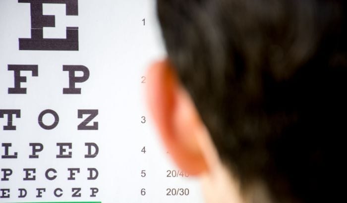
Visual acuity measures how well the visual system can detect and differentiate details (letters, edges, and contours) at a standardized distance. The test determines the smallest letter or symbol a person can correctly identify, with each eye tested separately.
The examination answers a straightforward question: at what minimum distance can the eye resolve a detail subtending one minute of arc? That concept, the minimum angle of resolution (MAR), is the foundation of every notation system described above.
High visual acuity does not equal perfect vision: it measures only central resolving power. Other components, including peripheral vision, color vision, and contrast sensitivity, require separate testing.
How the Snellen chart test is performed
In 1862, Dutch ophthalmologist Herman Snellen introduced a standardized system for assessing visual acuity using optotypes (characters drawn on a 5×5 unit grid), replacing the arbitrary reading passages that clinics had been using. The system made consistent comparisons between patients and institutions possible for the first time.
The chart consists of rows of letters that decrease progressively in size. The test is performed at 20 feet (6 meters in the metric system). The patient covers one eye and reads the letters from top to bottom; the last row read correctly defines their visual acuity for that eye. For how to convert that result to decimal or logMAR, see the conversion table above.
What the numbers mean: normal vision, low vision, and legal blindness
The World Health Organization (WHO) classifies visual impairment based on presenting visual acuity, meaning the acuity a person has with whatever correction they use in daily life, not under ideal clinic conditions. The definitions follow the ICD-11 (International Classification of Diseases, 11th Revision):
| Category | Visual acuity (better eye) |
|---|---|
| No visual impairment | 6/12 or better (≥ 20/40) |
| Mild impairment | Worse than 6/12 to 6/18 |
| Moderate impairment | Worse than 6/18 to 6/60 |
| Severe impairment | Worse than 6/60 to 3/60 |
| Blindness | Worse than 3/60, or visual field less than 10° |
According to the WHO report “Prevention of avoidable blindness and visual impairment” (EB124/7), blindness is defined as presenting visual acuity worse than 3/60 in the better eye, or a visual field of less than 10°.
What does 20/20 vision mean?
20/20 vision is not synonymous with perfect vision. It is the statistical reference standard described above: the eye resolves details that subtend 1 minute of arc, close to the theoretical resolving limit of the human eye. Some people achieve 20/15 or even 20/10, indicating above-standard acuity.
What does “low visual acuity” mean?
Low vision is the clinical term for visual acuity below 6/18 in the better eye, even with the best available optical correction. It differs from blindness: people with low vision can still see, but with significant functional limitation for everyday activities such as reading and recognizing faces.
What is legal blindness?
As explained above, the United States defines legal blindness through the Social Security Administration as best-corrected visual acuity of 20/200 or worse in the better eye, or a visual field of 20 degrees or less. The WHO threshold for blindness (worse than 3/60, equivalent to a 0.05 decimal score) is stricter than the U.S. threshold of 20/200 (0.1 decimal), and is used in global epidemiological surveys rather than U.S. disability determinations.
Main conditions that reduce visual acuity
Several eye conditions directly affect the sharpness of vision. The most common are:
Refractive errors
These account for the majority of cases of reduced visual acuity and are usually correctable with lenses.
- Myopia (nearsightedness): the eye focuses the image in front of the retina, making distant objects blurry. Corrected with diverging (minus) lenses.
- Hyperopia (farsightedness): the eye focuses the image behind the retina, causing difficulty with near focus (and in more pronounced cases, distance as well). Corrected with converging (plus) lenses.
- Astigmatism: an irregularity in the curvature of the cornea or lens distorts the image at all distances. Corrected with cylindrical lenses.
- Presbyopia: age-related decline in near-focusing ability from a loss of accommodation rather than a true refractive error, typically beginning around age 40. Corrected with multifocal or reading lenses.
When myopia and astigmatism occur together, a common combination, the correction approach differs from either condition alone. See: Myopia and Astigmatism: Differences, Symptoms, and Correction
Cataract
A cataract is the progressive clouding of the crystalline lens, the eye’s natural lens. As the lens loses transparency, the amount and quality of light reaching the retina decrease, gradually reducing visual acuity. Early symptoms include “foggy” vision, increased light sensitivity, and frequent changes in prescription. Treatment is surgical, replacing the clouded lens with an intraocular lens (IOL).
Age-related macular degeneration (AMD)
AMD affects the macula, the central region of the retina responsible for fine-detail vision. It causes progressive loss of central vision (the part of the visual field used for reading, recognizing faces, and performing precision tasks) while peripheral vision is usually preserved. There are two forms: dry (atrophy of retinal cells) and wet (abnormal blood vessel growth causing bleeding and edema).
Glaucoma
Glaucoma damages the optic nerve, usually due to elevated intraocular pressure. It initially affects peripheral vision, but without treatment it can progress to compromise central acuity. Because it is often silent in its early stages, early detection depends on routine eye examinations.
Visual acuity development in children
Visual acuity is not fully developed at birth. In newborns, the visual system is still immature, and visual sharpness improves progressively over the first years of life as the visual cortex matures.
Research published in PMC (Visual Acuity Norms in Preschool Children: The Multi-Ethnic Pediatric Eye Disease Study) shows that the proportion of children with acuity of 20/40 or better rises from 81% at 30–35 months to virtually 100% by 60–72 months.
Amblyopia: when development goes wrong
Amblyopia (“lazy eye”) occurs when the brain begins to suppress the visual input from one eye, usually because of strabismus, a significant difference in prescription between the eyes, or a visual obstruction during childhood. It is the most common cause of monocular visual impairment in children, with a prevalence of 2–3% in the pediatric population according to a review published in PMC (Amblyopia in children, 2014).
Treatment is most effective when started during the critical period of visual development, before age 7. Early vision screening, especially between ages 3 and 5, is essential to detect the condition in time.
Children rarely complain of poor vision in one eye because the other eye compensates. The only way to detect amblyopia is through a clinical examination that tests each eye separately.
When to have a visual acuity test
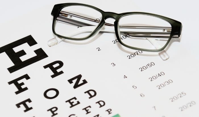
A visual acuity test is recommended:
- In early childhood: vision screening between ages 3 and 5 is recommended by the American Academy of Pediatrics and the U.S. Preventive Services Task Force to detect amblyopia and refractive errors early
- During school age: difficulty reading, trouble seeing the board, or habitually holding objects too close are warning signs
- In adulthood: every two years for adults without symptoms or risk factors; annually for those over 60 or with diabetes
- Whenever symptoms arise: sudden change in visual clarity, double vision, difficulty adjusting to light or darkness (including photophobia), or spots in the visual field
If you notice that your eyeglass prescription changes frequently or that your current lenses no longer fully correct your vision, a new acuity test can identify changes in your refraction. For help interpreting the numbers on the prescription itself, see how to read your prescription.
Warning signs that call for urgent eye care
Seek immediate attention if you or your child experience:
- Sudden onset of floaters accompanied by flashes of light (a possible sign of retinal detachment)
- Rapid loss of vision in one or both eyes
- Severe eye pain with reduced vision
- Sudden onset of double vision
- A “shadow” or progressively darkening area in the visual field
Who performs the visual acuity test
The test can be performed by:
- Ophthalmologist: a physician specializing in eye diseases and surgery; performs a comprehensive exam, including a dilated fundus examination
- Optometrist: a doctor of optometry licensed to perform refractions, prescribe corrective lenses, diagnose eye conditions, and in most U.S. states, treat certain eye diseases
- Optician: a trained professional who fits and dispenses eyeglasses and contact lenses based on a prescription; may perform basic acuity screening
- School nurse or trained screener: in school and community health screening programs
Prescribing corrective lenses or any therapeutic intervention requires evaluation by an ophthalmologist or optometrist.
When an acuity result should trigger a referral
An optician performing basic acuity screening is not diagnosing the cause of a reduced result, but a quick pinhole test can help decide whether a case needs a referral before ordering new lenses. According to the American Academy of Ophthalmology’s EyeWiki resource on visual acuity testing, if acuity improves when the patient looks through a pinhole occluder, the reduction is likely refractive, and a change in prescription should resolve it. If it does not improve, documented in clinical notes as “NI,” the cause is more likely something a lens correction will not fix on its own, such as a cataract or a retinal problem, and the patient should be referred to an optometrist or ophthalmologist rather than dispensed glasses based on the unimproved number. The same applies whenever a recorded result has dropped noticeably from a patient’s previous measurement without an obvious refractive explanation.
Telehealth and remote screening
In underserved areas with limited access to eye care specialists, telehealth models allow a trained technician to perform the visual acuity test at a clinic or school while an ophthalmologist or optometrist reviews the results and issues a report remotely. This workflow extends the reach of vision screening without compromising diagnostic quality.
Optogrid supports this model by enabling accurate digital measurement of optical parameters, such as pupillary distance, from a photograph, with no physical equipment required on-site.
Other tests that complement the visual acuity exam
The Snellen test evaluates central acuity only. For a complete visual assessment, an eye care provider may also perform:
- Ishihara test: 38 cards with colored circles containing numbers; detects and classifies color vision deficiency (absence or dysfunction of cone photoreceptors sensitive to specific colors)
- Potential Acuity Meter (PAM): uses a laser to assess retinal function directly, commonly indicated before and after eye surgeries such as cataract removal or corneal transplant
- Visual field testing (perimetry): maps the visual field, essential for detecting glaucoma
- Tonometry: measures intraocular pressure
- Optical coherence tomography (OCT): high-resolution imaging of the retinal layers
Understanding the anatomy of the human eye helps clarify why each of these tests targets a different structure or function.
Frequently asked questions about visual acuity
What is visual acuity?
Visual acuity measures how sharply the eye can resolve fine detail at a standardized distance. The result indicates the smallest line of letters or symbols a person can identify on the Snellen chart. For the four notations used to report the result, see the conversion table above.
What does 6/12 vision mean?
6/12 is the same result as 20/40, decimal 0.5, or logMAR 0.3. It is a mild reduction that still falls within the WHO’s “no visual impairment” category (6/12 or better), and it usually reflects an uncorrected or under corrected refractive error rather than a serious eye condition.
What does 0.1 visual acuity mean?
A decimal score of 0.1 is the same as 6/60 or 20/200. In the United States, 20/200 or worse in the better eye is the Social Security Administration’s threshold for legal blindness. On the WHO scale, 6/60 marks the boundary between moderate and severe visual impairment.
How is the visual acuity test done?
The patient stands 20 feet (6 meters) from the Snellen chart, covers one eye, and reads the letters or symbols from top to bottom. The last row read correctly defines the acuity for that eye. The test is non-invasive and takes only a few minutes.
What is the difference between Snellen, decimal, and logMAR scales?
Snellen expresses acuity as a fraction (20/20, 20/40); decimal reduces that same fraction to a single number (1.0, 0.5); logMAR expresses it as the logarithm of the decimal’s reciprocal (0.0, 0.3). LogMAR is more precise for clinical research and patient follow-up because its increments are uniform. All three describe the same visual capability, just in different units.
What is legal blindness?
In the United States, legal blindness is defined as best-corrected visual acuity of 20/200 or worse in the better eye (0.1 decimal, 6/60 metric), or a visual field of 20 degrees or less. The WHO uses a stricter threshold for blindness: presenting visual acuity worse than 3/60 in the better eye, or a visual field of less than 10°.
When do children reach adult-level visual acuity?
Most children reach 20/20 acuity between ages 5 and 7. Visual development begins at birth and progresses rapidly during the first three years. That is why vision screening between ages 3 and 5 is critical for detecting conditions like amblyopia before the critical developmental window closes.
Who can prescribe glasses after a visual acuity test?
In the United States, prescribing corrective lenses is within the scope of practice of ophthalmologists and optometrists. Opticians fit and dispense eyewear based on a valid prescription but do not prescribe. For progressive lenses, bifocals, or cases involving ocular disease, evaluation by an ophthalmologist or optometrist is required.
References
- Messias A, Jorge R, Cruz AA. Tabelas para medir acuidade visual com escala logarítmica: porque usar e como construir [Tables for measuring visual acuity on a logarithmic scale]. Arquivos Brasileiros de Oftalmologia. 2010;73(1):96–100. SciELO Brasil
- Social Security Administration. Statutory Blindness, POMS DI 26001.001. secure.ssa.gov
- Snellen H. Optotypi ad visum determinandum. Utrecht: P.W. van de Weijer, 1862. Historical context: PubMed: Snellen and his optotypes
- World Health Organization. Prevention of avoidable blindness and visual impairment. Document EB124/7. Geneva: WHO, 2009. WHO
- World Health Organization. Blindness and visual impairment: Fact sheet. who.int
- Donahue SP, Nixon CN; Section on Ophthalmology, American Academy of Pediatrics. Visual system assessment in infants, children, and young adults by pediatricians. Pediatrics. 2016;137(1). Preschool acuity norms: PMC Visual Acuity Norms in Preschool Children
- Webber AL, Wood JM. Amblyopia: prevalence, natural history, functional effects and treatment. Clinical and Experimental Optometry. 2005. Pediatric amblyopia review: PMC Amblyopia in children
- American Academy of Ophthalmology. Visual Acuity Testing in Adults. EyeWiki. eyewiki.aao.org

I grew up inside an optical shop. My mother likes to tell how, as a kid, I would watch eyeglasses being assembled, play with the tools, and draw on the back of service order slips. The family business taught me early what a precise measurement and work done right are worth.
I went on to spend more than twenty years as a software engineer, always keeping an eye on the optical world. Optogrid was born where the two meet: digital measurement technology (pupillary distance and fitting heights) built by someone who knows the dispensing counter from the inside.