
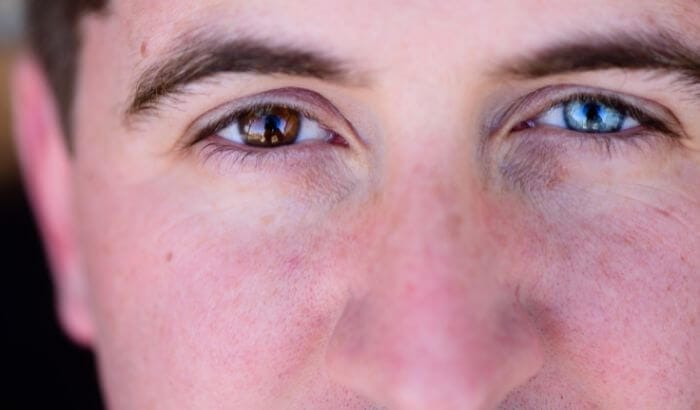
Heterochromia is a rare condition in which a person has differently colored eyes, or color variation within the same iris. Scientific studies indicate it affects roughly 0.06% of the general population, making it far more common in animals than in humans. The three main forms are complete (each eye a different color), sectoral (a patch within one iris), and central (a contrasting ring around the pupil). In the vast majority of cases, it is a benign genetic trait that does not affect vision or pose any risk to eye health.
Have you ever had a customer walk into your practice and, when you looked directly at them, you noticed that one eye was a completely different color, or that one iris had a distinct patch that set it apart from the rest? That is heterochromia: a rare condition that gives some people two different eye colors, or a striking variation of color within a single iris.
It is a characteristic that sparks curiosity and, understandably, raises questions about its origins and what it means for a person’s eye health. This guide covers everything an eye care professional needs to know: what heterochromia is, its three types, the genetic and acquired causes, when it signals a problem, and the most persistent myths surrounding it.
What Is Heterochromia?
Heterochromia (or heterochromia iridis) is a variation in eye color that occurs either between the two eyes or within different areas of a single eye. The difference comes down to variation in the amount and distribution of melanin, the pigment responsible for color in the skin, hair, and eyes.
According to the Cleveland Clinic, heterochromia encompasses three types: in complete heterochromia, “one iris is a different color than the other”; in sectoral heterochromia, “one iris has a section that’s a different color from the rest”; and in central heterochromia, “one iris has an inner ring that’s a different color from the rest.”
In most people, melanin is distributed evenly across both irises. In people with heterochromia, that distribution differs, resulting in eyes with distinct color patterns:
- Between the two eyes (one blue, one brown, for example)
- Within the same eye (an iris with two clearly distinct color zones)
- Around the pupil (a ring of contrasting color near the center)
How Melanin Determines Eye Color
Eye color is directly related to the amount of melanin in the front layers of the iris. People with brown eyes have a large amount of melanin in the iris, while people with blue eyes have much less of this pigment.
The OCA2 and HERC2 Genes
A specific region on chromosome 15 plays a central role in determining eye color, and it contains two closely positioned genes: OCA2 and HERC2. The protein produced by OCA2 is involved in the maturation of melanosomes, the cellular structures that produce and store melanin.
The HERC2 gene contains a region called intron 86, which holds a segment of DNA that controls the activity of the OCA2 gene, turning it on or off as needed. Less OCA2 expression means less melanin in the iris, producing blue eyes rather than brown.
In heterochromia, this melanin distribution is irregular or asymmetric, which creates the distinctive color patterns described below.
3 Types of Heterochromia: Complete, Sectoral, and Central
There are three main types of heterochromia, each with distinct visual characteristics. Understanding the differences is useful for opticians and ophthalmologists when documenting a patient’s ocular characteristics.
Comparison Table: Types of Heterochromia
| Type | Defining Feature | Visual Example | Relative Frequency |
|---|---|---|---|
| Complete | Each eye is a completely different color | One blue eye, one brown eye | Least common |
| Sectoral (Partial) | A segment of one iris differs from the rest | Brown iris with a blue or green sector | More common |
| Central | A ring of contrasting color surrounds the pupil | Golden/green ring near pupil, blue toward the outer iris | Most frequent |
Complete Heterochromia
This is the most visually striking type: each eye is a distinctly different color. One eye might be blue and the other brown, creating an unmistakable appearance. Research analyzing 11,111 high-resolution yearbook portraits found only 7 confirmed cases, placing the observed prevalence of complete heterochromia at 0.063%, with a 95% confidence interval of 0.028% to 0.133%.
Sectoral Heterochromia (Partial)
In sectoral heterochromia, one region of an iris is a different color from the rest. This happens when melanin concentrates unevenly in just one area of the iris.
According to research on congenital sectoral heterochromia, “in sectoral heterochromia, part of one iris is a different color from its remainder”, and the pattern can affect one or both eyes. A brown iris might have a region that appears blue or greenish, almost like a birthmark of color. The arrangement can follow a radial pattern, giving each case its own signature appearance.
Central Heterochromia
Here, color variation appears as a ring around the pupil, typically a contrasting inner zone that gradually gives way to a different color toward the outer edge of the iris. The effect is often subtle and most visible under good lighting, but it is the most frequently occurring type of heterochromia.
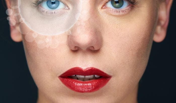
Why Do Some People Have Different Colored Eyes? Genetic and Acquired Causes
Heterochromia can be congenital (present from birth) or acquired (developing at some point during life). The two origins have different characteristics and very different implications for health.
Congenital Heterochromia: Genetic Factors
Genetics is the main driver of congenital heterochromia. It can run in families, transmitted through autosomal dominant inheritance, with documented cases including families where the trait appeared across three consecutive generations.
Primary genetic causes:
- Benign genetic mutation: Many cases result from a mutation that affects only melanin distribution in the iris, without impacting eye anatomy or function.
- Genetic mosaicism: A mutation during cell division (mitosis) can produce cells with different levels of melanin in different parts of the body, including the iris.
- Chimerism: A rare condition in which a person carries two distinct sets of DNA, which can manifest as pigmentation differences across the body, including the eyes.
In the vast majority of cases, congenital heterochromia is completely harmless. Many people go years without noticing it themselves until someone else points it out.
A note from practice: At the dispensing counter, subtle central heterochromia often goes completely unnoticed by patients. Under the focused lighting used when fitting frames, an inner ring of contrasting color becomes visible in a way that ordinary mirrors do not reveal. More than once, a patient has been surprised when an optician mentioned it during a frame fitting, having lived for decades unaware the variation was there at all. That moment of curiosity, handled with a calm factual explanation, almost always turns into a positive interaction.
Acquired Heterochromia: Causes During Life
Acquired heterochromia develops later in life due to a range of factors:
Main causes:
- Eye trauma: Injuries that disrupt melanin production or distribution in the iris
- Eye diseases: Glaucoma, uveitis, intraocular tumors
- Medications: Certain eye drops, particularly prostaglandin analogs used for glaucoma, can darken iris pigmentation over time
- Eye surgery: Procedures that alter iris structure
- Systemic conditions: Some diseases affect ocular pigmentation over time
Important note for practitioners: Heterochromia that appears suddenly in an adult should always be evaluated by an ophthalmologist, as it may indicate an underlying condition requiring treatment.
Medical Conditions Associated with Heterochromia
While congenital heterochromia is generally benign, it can be associated with specific medical conditions worth recognizing in clinical practice.
Waardenburg Syndrome
Waardenburg syndrome is a rare genetic condition with an estimated global prevalence of approximately 1 in 42,000 individuals. It is present in 0.9% to 2.8% of people with congenital hearing loss.
Key features:
- Heterochromia (complete or sectoral) or brilliantly blue irises in both eyes
- White forelock (poliosis)
- Broad nasal root
- Congenital sensorineural hearing loss
- Telecanthus (increased distance between the inner corners of the eyes)
Heterochromia is one of the defining characteristics of Waardenburg Type 1, the most common form of the syndrome.
Other Associated Conditions
- Horner syndrome: Can cause heterochromia when it occurs in infants (the affected eye develops less pigmentation)
- Fuchs heterochromic cyclitis: An inflammatory condition that can cause gradual lightening of the affected iris
- Iris melanoma: A tumor that can alter iris pigmentation (rare)
- Ocular siderosis: Iron deposition in the eye following trauma
How Common Is Heterochromia? Prevalence Data
Heterochromia is genuinely rare in humans. The most robust evidence comes from two independent studies that arrived at nearly identical estimates despite being conducted decades apart and in different populations.
Prevalence in Humans
- Complete heterochromia: Approximately 0.063% of the general population, roughly 6 in every 10,000 people
- All types combined: Less than 1% of the world population
- Historical benchmark: Stelzer’s Vienna study of over 25,000 residents found a prevalence of 0.063%, virtually identical to the modern yearbook study, which the authors note: “Our point estimate is nearly identical to Stelzer’s result, and our CI contains it.”
Prevalence in Animals: Much Higher
Heterochromia is significantly more common in domestic animals:
In cats:
- Particularly frequent in white cats and in breeds such as Turkish Van
- Certain dog breeds with color-dilution genes have substantially elevated rates
In dogs:
- Australian Shepherds, Dalmatians, and Siberian Huskies are among the breeds most commonly affected
Why is it more common in animals?
The answer lies in selective breeding: when animals from the same litter share a gene for heterochromia and later mate, their offspring have a higher probability of carrying the trait forward. Breeds with color-dilution genes or genes associated with white coats tend to show the highest rates.
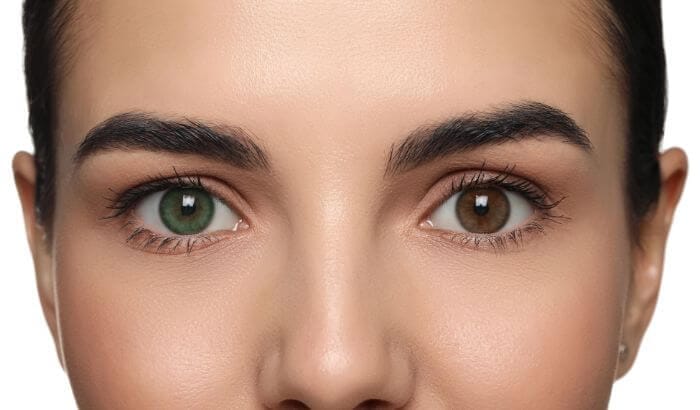
Heterochromia in Famous People: Myths and Facts
Several celebrities are known for their unusual eye coloring, but not all of them actually have heterochromia, and this is where a common and persistent misconception lives.
David Bowie: The Most Famous Case (and the Most Misunderstood)
Many people believe David Bowie had heterochromia. This is incorrect. Bowie had anisocoria, a condition in which the pupils are different sizes, not different eye colors. The American Academy of Ophthalmology distinguishes clearly between the two: heterochromia is a difference in iris pigmentation, while anisocoria is a difference in pupil size.
What actually happened:
- In February 1962, Bowie was punched in the left eye by his friend George Underwood during a dispute (Bowie Bible, 12 February 1962)
- The injury resulted in four months of hospital treatment and left his left pupil permanently dilated
- The permanently dilated pupil made his left iris appear much darker than his right, creating the illusion of differently colored eyes
- Both eyes were actually the same color. The visual difference came entirely from the dilated pupil, not from different levels of iris pigmentation
Remarkably, Bowie reportedly thanked Underwood for the injury that became one of his most recognizable features, and the two remained friends throughout their lives.
Celebrities with True Heterochromia
- Kate Bosworth: Sectoral heterochromia, her right eye is partially blue and partially brown
- Mila Kunis: Complete heterochromia (one green eye, one brown)
- Max Scherzer (MLB pitcher): Complete heterochromia (one blue eye, one brown)
- Alice Eve (actress): Complete heterochromia
Myths and Facts About Heterochromia
Myth 1: Heterochromia always indicates a disease
Fact: The large majority of congenital heterochromia cases are completely benign and unrelated to any medical condition. It is simply a natural variation in melanin distribution.
Myth 2: Heterochromia affects vision
Fact: Heterochromia itself does not affect visual acuity or the quality of sight. The color variation in the iris has no bearing on how the eye functions optically. People with heterochromia see just as well as anyone else.
Myth 3: Heterochromia can be “cured” or reversed
Fact: Congenital heterochromia is a permanent genetic characteristic, not a disease requiring correction. There is no treatment for it, nor any reason to look for one. Many people with heterochromia consider it a distinctive personal feature.
Myth 4: Only people with light eyes can have heterochromia
Fact: Heterochromia can occur at any baseline eye color. There can be heterochromia between a hazel eye and a green eye, or between lighter and darker shades of brown.
Fact 1: Heterochromia can be hereditary
True. Cases of sectoral heterochromia with autosomal dominant inheritance have been documented in families across three generations. That said, many cases are sporadic, the result of random mutation or mosaicism rather than an inherited trait.
Fact 2: Sudden heterochromia in adults requires evaluation
True. If heterochromia appears suddenly or is accompanied by other symptoms (pain, visual changes, redness), a prompt consultation with an ophthalmologist is warranted to rule out secondary causes.
Fact 3: Animals have heterochromia at far higher rates than humans
True. While the condition affects less than 1% of humans, it is considerably more common in certain animal populations due to selective breeding practices.
When to Worry: Heterochromia and Eye Health
Patients, and their families, often want to know whether heterochromia puts their eyes at risk. The answer depends on the type and timing of onset.
Congenital Heterochromia: Generally No Risk
The majority of cases present from birth and remain completely stable throughout life. If a person has always had differently colored eyes and has no other symptoms, there is no clinical concern.
Staff training note: When a new patient presents with heterochromia, a brief intake question, “Has your eye color always been like this?” takes seconds and has real clinical value. A “yes, since birth” answer points to congenital heterochromia, which needs no urgent action. Any answer involving recent change warrants a same-day note to the examining optometrist or ophthalmologist.
Warning Signs: When to Refer to an Ophthalmologist
Recommend a specialist consultation if any of the following apply:
- Heterochromia that develops suddenly in adulthood
- Progressive change in iris color over weeks or months
- Accompanying symptoms:- Eye pain – Blurred vision – Light sensitivity – Persistent redness – Any degree of vision loss
- Recent eye trauma
- New systemic medications, particularly glaucoma eye drops
Follow-Up for Acquired Heterochromia
When the cause is acquired, ongoing monitoring is standard practice. Depending on the suspected cause, an ophthalmologist may order:
- Detailed slit-lamp biomicroscopy
- Tonometry (intraocular pressure measurement)
- Imaging (if tumor or structural cause is suspected)
- Genetic evaluation (in syndromic presentations)
FAQ: Common Questions About Heterochromia
1. Can heterochromia change over time?
Congenital heterochromia generally remains stable throughout life. That said, eye color can undergo subtle shifts during infancy and early childhood as melanin deposits in the iris mature. Significant iris color changes in adults should be evaluated by an ophthalmologist.
2. Is heterochromia hereditary? Will parents with heterochromia pass it to their children?
It can be. Sectoral heterochromia in particular has been documented with autosomal dominant inheritance across multiple generations. However, many cases arise sporadically from random mutations or mosaicism, so having heterochromia does not guarantee passing it on.
3. Do people with heterochromia need special eye care?
No. Congenital heterochromia does not require any specific clinical management beyond standard eye health recommendations: regular eye exams, UV protection, and precise pupillary distance measurements when fitting eyeglasses.
4. Do colored contact lenses work normally on eyes with heterochromia?
Yes. Colored contact lenses function normally regardless of iris pigmentation. Some people with heterochromia choose tinted lenses to unify the appearance of their eyes; others prefer to keep their natural variation.
5. Can heterochromia affect color vision or color perception?
Heterochromia does not affect color vision or the ability to distinguish between colors. Color perception depends on the photoreceptors (cones) in the retina, not on iris pigmentation. A person with two differently colored irises perceives color the same way as anyone else. If color vision concerns arise, a dedicated color vision test can assess cone function independently.
6. Can babies be born with heterochromia?
Yes. Congenital heterochromia is present from birth. One important note: many newborns have light-colored eyes that deepen over the first few months as melanin deposits in the iris. True heterochromia will be apparent once the iris color has stabilized, usually by around six to twelve months of age.
7. What is the difference between heterochromia and anisocoria?
These two conditions are frequently confused. Heterochromia refers to differences in iris color, the pigmented ring around the pupil. Anisocoria refers to unequal pupil sizes. David Bowie is the classic example: his eyes appeared different in color because one pupil was permanently dilated from a February 1962 injury, not because his irises contained different amounts of melanin.
8. Why is heterochromia more common in certain dog and cat breeds?
The elevated frequency in certain breeds comes down to selective breeding and limited gene pools. When animals from the same litter, sharing the same genetic variants for pigmentation, reproduce together, offspring are more likely to inherit those same traits. Breeds with color-dilution genes or genes associated with white coats tend to show the highest rates.
Fitting Eyewear for Patients with Heterochromia
Patients with heterochromia are not a clinical exception, but they can benefit from a more considered conversation at the dispensing counter. Their distinctive eye coloring has no bearing on the measurements required to produce well-fitting, optically accurate eyeglasses, but frame aesthetics are another matter.
A few practical considerations worth raising:
- Frame color and iris contrast: Patients with complete heterochromia sometimes prefer frame colors that complement their lighter eye rather than compete with it. A warm tortoise frame, for example, can pull together a hazel-brown and blue combination without drawing unequal attention to either eye.
- Lens tint and photochromic lenses: Both eyes adapt identically to light regardless of iris pigmentation, so there is no optical reason to avoid tinted or photochromic lenses. Some patients with central heterochromia appreciate how certain tints reduce the visibility of the inner ring variation, if self-consciousness is a concern.
- Mirror coatings on sunglasses: Patients who are self-conscious about their heterochromia often appreciate that mirrored or solid-tint sunglass lenses eliminate the visible difference entirely when they are outdoors.
- The consultation itself: Simply explaining what heterochromia is, and confirming it poses no health risk, is often the most valuable thing an optician can offer. Many patients, particularly those newly diagnosed as adults, arrive with anxiety about what the color difference means. A confident, evidence-grounded explanation at the dispensing chair is both a clinical service and a practice differentiator.
How Optogrid Helps with Customers Who Have Heterochromia
Customers with heterochromia deserve the same precise, attentive fitting process as every other patient. Their distinctive eye coloring has no bearing on the measurements required to produce well-fitting, optically accurate eyeglasses.
Accurate Measurements Regardless of Iris Color
The correct pupillary distance (PD) is critical to lens comfort and optical performance. Optogrid measures monocular and binocular PD directly from a photo, in a matter of minutes, regardless of eye color or iris pattern.
What Optogrid does:
- Measures monocular and binocular PD from an image with a known reference (card, frame width, ruler)
- Calculates fitting height and records measurements visually for easy review
- Optician workflow: a secure link lets the customer send their own photo remotely; measurements are stored in a history panel for verification
- Runs in the browser with no installation required, compatible with e-commerce and in-store sales workflows
- Precision: measurements to 0.5 mm, ensuring optimal visual comfort
Serving Every Customer Well
Having two differently colored eyes is a distinctive personal feature, not a clinical limitation. Your practice can turn that moment of curiosity into a high-quality consultation: offering accurate measurements, answering questions with confidence grounded in science, and suggesting frames that complement rather than compete with what makes each customer unique.
Ready to improve your measurements and deliver exceptional care to every customer?
Try Optogrid for free and see how precise, photo-based measurements make a difference for every customer who walks through your door.
Sources and Scientific References
- Estimating the prevalence of heterochromia iridum from high-resolution digital yearbook portraits. PMC / National Library of Medicine
- Heterochromia, StatPearls Medical Library. NCBI Bookshelf
- Artistic Iris: A Case of Congenital Sectoral Heterochromia Iridis. PMC / National Library of Medicine
- Is eye color determined by genetics?. MedlinePlus Genetics
- Association between Variants in the OCA2-HERC2 Region and Blue Eye Colour. PMC / National Library of Medicine
- Waardenburg Syndrome, StatPearls. NCBI Bookshelf
- Heterochromia: Types, Causes, and When to Seek Care. Cleveland Clinic
- What Is Heterochromia?. American Academy of Ophthalmology
- George Underwood Punches David Bowie. Bowie Bible (12 February 1962)

I grew up inside an optical shop. My mother likes to tell how, as a kid, I would watch eyeglasses being assembled, play with the tools, and draw on the back of service order slips. The family business taught me early what a precise measurement and work done right are worth.
I went on to spend more than twenty years as a software engineer, always keeping an eye on the optical world. Optogrid was born where the two meet: digital measurement technology (pupillary distance and fitting heights) built by someone who knows the dispensing counter from the inside.