

The Ishihara color plate test is the most widely used color blindness screening method. Its sensitivity reaches 99% at a three-error threshold, according to research in Ophthalmic and Physiological Optics. For general office screening, Ishihara plates are sufficient. Precise classification of the type and severity requires more than a screening test. For occupational certification or driving fitness, clinicians use the Nagel anomaloscope or the Farnsworth D-15 arrangement test. Anyone having trouble telling colors apart should see an ophthalmologist for a formal evaluation.
What Color Blindness Is and How It Affects Vision
Color blindness, clinically called color vision deficiency (CVD), reduces or removes the ability to tell certain colors apart. It results from anomalies in the retinal cones, the light-sensitive cells that sense color. Each cone type responds to a different wavelength of light: red (L-cones), green (M-cones), and blue (S-cones). When one or more cone types are missing or work abnormally, color vision deficiency occurs.
The condition is mostly hereditary and X-linked, which explains the sharp prevalence difference between sexes. Research published in the Journal of the Optical Society of America (Birch, 2012) studied red-green color deficiency in European Caucasians. It found a prevalence of “about 8% in men and about 0.4% in women”. The National Eye Institute states that “about 1 in 12 men have color vision deficiency”. This confirms that men face much higher risk than women.
Color vision deficiency can also be acquired later in life. Causes include glaucoma, diabetic retinopathy, multiple sclerosis, or exposure to certain toxins and medications. Acquired CVD may affect one or both eyes unevenly, and it tends to worsen over time if the underlying condition goes untreated.
Clinical Classification of Color Vision Deficiency Types
Color blindness is classified by the number of functional cone types and which cone is affected.
Anomalous trichromacy (three cones, one malfunctioning)
This is the most common form. All three cone types are present, but one responds abnormally.
- Protanomaly: L-cones (red) have reduced sensitivity. Red appears dimmer and is often confused with green or brown.
- Deuteranomaly: M-cones (green) function abnormally. This is the most prevalent form of color vision deficiency overall. Green is perceived differently and confused with red or yellow.
- Tritanomaly: S-cones (blue) are deficient. Blue and green appear similar; yellow and violet may be confused. This form is rare.
Dichromacy (two functional cones, one absent)
One of the three cone types is completely absent. This leaves only two photoreceptor types for color perception.
- Protanopia: absence of L-cones. Red is perceived as black or dark gray. Distinguishing any color containing red becomes difficult.
- Deuteranopia: absence of M-cones. Green, red, and yellow become indistinguishable in many combinations.
- Tritanopia: absence of S-cones. Blue and green are confused; yellow and violet as well. Rare, and distributed equally between men and women because it is not X-linked.
Monochromacy (achromatopsia)
The rarest and most severe form. The person sees only in shades of gray because all cones are absent or nonfunctional. Achromatopsia has an estimated prevalence of 1 in 30,000. It often involves photophobia (intense light sensitivity) and reduced visual acuity.
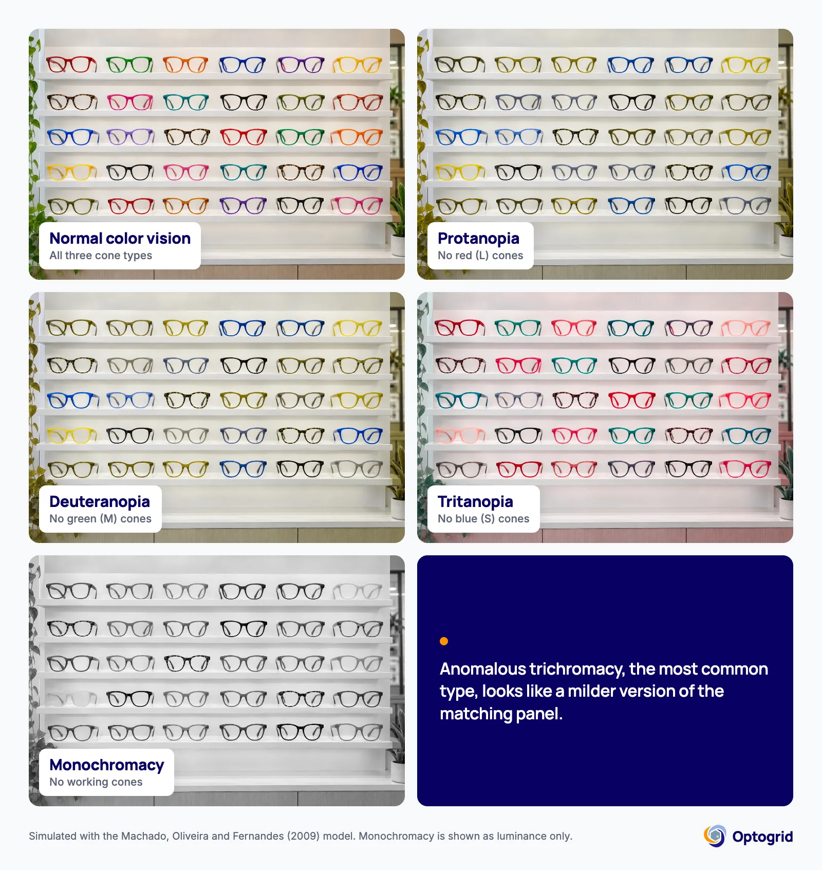
The simulation below shows the same optical shop frame display through each type of dichromacy and through monochromacy. With protanopia and deuteranopia, red and green frames merge into the same olive tones. With tritanopia, blue and green are confused and yellow turns pink.
Four Color Blindness Tests Compared: Methods, Strengths, and Limits
Several diagnostic methods exist for color vision deficiency, each suited to a different clinical purpose. The table below summarizes how they compare before examining each test in detail.
| Test | Method | Best For | Sensitivity | Limitations |
|---|---|---|---|---|
| Ishihara plates | Pseudoisochromatic dot patterns with hidden numbers | Red-green screening | Up to 99% (3-error threshold) | Does not detect tritan defects; no severity grading. |
| Nagel anomaloscope | Color-matching with adjustable red-green light mixture | Definitive diagnosis and classification | Gold standard | Expensive, requires trained operator, time-consuming. |
| Farnsworth D-15 | Arrangement of 15 colored caps in sequence | Occupational fitness; separating severe from mild | Good for dichromacy detection | Less sensitive to mild anomalous trichromacy. |
| Holmgren wool test | Sorting colored wool skeins by hue | Basic screening | Low | Largely replaced by plate tests. |
Screening and diagnosis answer different questions. A screening test like Ishihara plates flags whether a deficiency is likely present. Diagnosis, meaning the exact type and severity, requires the Nagel anomaloscope or a full clinical exam. A clean screening result does not rule out a mild deficiency. A positive one is not a final diagnosis on its own.
Ishihara Color Plate Test
Japanese ophthalmologist Shinobu Ishihara created the test in 1918 for military screening. It remains the most widely used color vision screening tool worldwide. It consists of 38 plates (with shorter 14- and 24-plate versions available) containing circles of colored dots at varying sizes and intensities. Dots of a different color form numbers or paths against the background. People with normal color vision read them clearly; people with a deficiency read them differently or not at all.
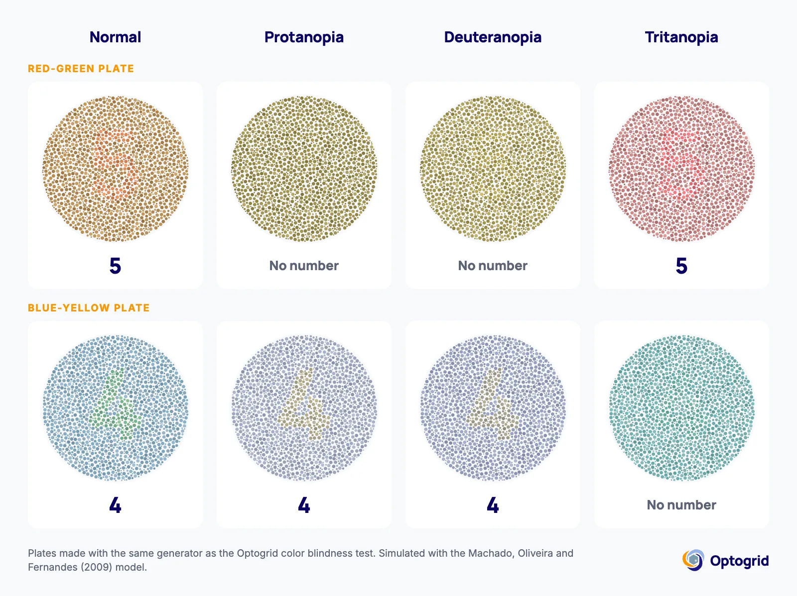
In the simulation below, made with the plate generator of Optogrid’s online color blindness test, the number in a red-green plate disappears with protanopia and deuteranopia, while a blue-yellow plate loses its number only with tritanopia.
The AAO describes the Ishihara as “one of the most common and reliable color blind tests” (AAO: How Color Blindness Is Tested). Research by Birch (1997) in Ophthalmic and Physiological Optics quantified its performance: “the combined sensitivity of the Transformation and Vanishing plates of the 38 plate Edition of the Ishihara plates is 95.5% on eight errors, 97.5% on six errors and 99.0% on three errors”.
Pseudoisochromatic plate tests overall detect about 96% of cases confirmed by anomaloscope. This is according to the National Research Council’s Procedures for Testing Color Vision.
Limitations: The Ishihara detects red-green deficiencies with high sensitivity. It is less effective at identifying blue-yellow deficiencies (tritanopia/tritanomaly) and cannot classify severity.
Nagel Anomaloscope
The Nagel anomaloscope is the gold standard for diagnosing and classifying color vision deficiencies. The instrument presents two luminous fields side by side. One field is fixed yellow. The other is adjustable, formed by a mixture of red and green light. The patient adjusts the red-green ratio until the adjustable field appears identical to the yellow reference.
The National Research Council states that “the anomaloscope is the only clinical instrument for diagnosis and classification” (NCBI Bookshelf). This applies to congenital X-linked color vision deficiencies. The width of the matching range accepted by the patient indicates the exact type (protanopia, deuteranopia, protanomaly, or deuteranomaly) and its severity.
Limitations: The anomaloscope is expensive and requires special training in color analysis principles to operate and interpret. The exam also takes significant time. For these reasons, anomaloscopy is reserved for specialized clinical settings or occupational certifications with strict visual requirements.
Farnsworth D-15 Arrangement Test
The Farnsworth D-15 (dichotomous) test consists of 15 colored caps that the patient arranges in sequence according to perceived color similarity. The expected sequence follows a defined chromatic order. Diagonal crossovers reveal the axis of color confusion and the severity of the deficiency.
The D-15 was designed as a dichotomous test. Its purpose is to separate people with severe deficiency from those with mild deficiency or normal vision. It is widely used in occupational assessments for pilots, emergency vehicle drivers, and operators of color-coded systems. It is quick and portable.
The desaturated version (Lanthony D-15) uses caps with lower saturation. It is more sensitive for detecting mild deficiencies. It also picks up acquired color vision loss caused by conditions such as glaucoma, diabetic retinopathy, or toxic exposure.
Holmgren Wool Test
The Holmgren test uses skeins of colored wool that the patient sorts by perceived color. It is simple and inexpensive, useful for basic screening. But it has lower diagnostic precision than plate tests and arrangement methods, and modern clinical practice has largely replaced it.
Who Should Take a Color Blindness Test
Color vision testing is indicated for several groups:
- School-age children: Difficulty distinguishing colors can interfere with learning through color-coded charts, maps, and diagrams. Screening before school entry helps identify children who may need accommodations.
- People with family history: Red-green color blindness is hereditary. Sons of carrier mothers have a 50% chance of being affected.
- Candidates for visually demanding occupations: Pilots, commercial drivers, military personnel, electricians, and surgeons frequently need formal color vision assessment. This is part of occupational fitness certification.
- Adults with new color confusion complaints: Acquired color vision deficiency can signal ocular disease (glaucoma, macular degeneration) or systemic conditions (diabetes, multiple sclerosis). These cases should be investigated by an ophthalmologist.
- Before procedures affecting retinal function: Surgeries or treatments with potential impact on the retina may warrant baseline color vision testing.
What Color Blindness Test Results Mean
Results vary depending on the test used.
Ishihara plates:
- Correct reading of all plates: normal color vision.
- Errors on transformation and vanishing plates: suspected red-green deficiency.
- Different error patterns: indicate the type of deficiency (protan or deutan).
- The test provides screening only, not severity grading.
Nagel anomaloscope:
- Narrow matching range centered on the normal point: normal color vision.
- Wide matching range: anomalous trichromacy (the wider the range, the more severe).
- Acceptance of any mixture (full range): confirmed dichromacy (protanopia or deuteranopia).
- Reduced brightness of the red field: indicates protanomaly or protanopia.
Farnsworth D-15:
- Sequence without crossovers: normal vision or mild deficiency.
- Diametrically opposite crossovers: moderate to severe deficiency. The crossover axis indicates the type (protan, deutan, or tritan).
A positive Ishihara screening should always be confirmed by a comprehensive evaluation. This matters most when the deficiency has occupational implications or appears for the first time in adulthood. Acquired color vision loss can indicate an underlying eye disease that requires treatment.
Color vision testing is one component of a complete visual assessment that also includes a visual acuity test and pupil function evaluation. Related refractive conditions are often identified during the same comprehensive exam, including astigmatism and cases where myopia and astigmatism occur together. Understanding your full eyeglasses prescription also helps you manage these related refractive conditions.
Treatment and Management: What Works and What Does Not
There is no cure for congenital color blindness. Available options focus on adaptation strategies and, in some cases, devices that improve color discrimination for certain tasks.
Color filter lenses (such as ChromaGen and EnChroma) alter how colors reach the eye. They increase contrast between red and green for some patients. However, a systematic review and meta-analysis published in Health Science Reports (2022) concluded that these devices “do not provide clinically significant evidence that subjective color perception has improved” (PubMed). The effects are modest, vary widely between individuals, and cease when the lenses are removed.
Color identification apps (available for iOS and Android) use the smartphone camera to name colors in real time. These are practical accessibility tools for daily life. They do not depend on clinical evidence of efficacy. They simply serve an assistive function.
Environmental and educational adaptations have the greatest proven impact on quality of life. Examples include labels and legends that do not rely solely on color, plus software with accessible palettes. Open communication with employers and schools about the condition also helps.
For acquired color vision deficiency caused by disease or medication, treating the underlying condition may stabilize or partially reverse the deficiency. Regular follow-up with an ophthalmologist is essential in these cases. Children with refractive errors alongside color vision deficiency benefit from early intervention, including appropriate myopia control strategies when applicable.
Frequently Asked Questions
What is the most accurate color blindness test?
The Nagel anomaloscope is the gold standard. The National Research Council describes it as “the only clinical instrument for diagnosis and classification” of congenital X-linked color vision deficiencies. It identifies the exact type and severity. The Ishihara test is highly sensitive for screening (up to 99%) but does not classify severity.
Can children be tested for color blindness?
Yes. Ishihara plates have versions with simple shapes (circles and paths) instead of numbers, designed for preschool-age children. International protocols recommend screening between ages 3 and 5. This detects reduced acuity and color deficiency before school entry, when color-coded materials become common in the classroom.
What is the difference between the Ishihara test and the Farnsworth D-15?
The Ishihara is a screening test: it identifies whether a red-green deficiency exists. The Farnsworth D-15 is an arrangement test that separates severe deficiencies from mild ones and reveals the axis of color confusion. The D-15 is more common in occupational assessments; the Ishihara is the standard for general clinic-based screening.
Does color blindness affect driving eligibility?
It depends on the jurisdiction and the severity of the deficiency. Many countries require color vision assessment as part of driver licensing, particularly for commercial vehicle operators. Severe dichromacy may restrict certain license categories. An ophthalmologist can determine fitness through formal testing.
Do EnChroma or ChromaGen glasses cure color blindness?
No. A 2022 systematic review in Health Science Reports concluded that commercially available color vision devices “do not provide clinically significant evidence that subjective color perception has improved.” These lenses may enhance contrast for some users, but the effect varies between individuals and stops when the glasses are removed.
Can color blindness develop later in life?
Yes. Acquired color vision deficiency can result from ocular diseases (glaucoma, diabetic retinopathy, macular degeneration) or systemic conditions (diabetes, multiple sclerosis). It can also follow medication side effects or toxic exposure. Unlike congenital CVD, acquired forms often affect one eye more than the other and may worsen progressively.
Which professional performs color blindness testing?
Optometrists and ophthalmologists both perform color vision tests. Optometrists typically conduct routine screening (Ishihara, D-15) as part of comprehensive eye exams. Ophthalmologists are indicated when acquired color vision loss from disease is suspected or when anomaloscope testing is needed for precise classification.
References
- Birch J. Worldwide prevalence of red-green color deficiency. Journal of the Optical Society of America A. 2012;29(3):313-320. PubMed PMID 22472762
- Birch J. Efficiency of the Ishihara test for identifying red-green colour deficiency. Ophthalmic and Physiological Optics. 1997;17(5):403-408. PubMed PMID 9390366
- American Academy of Ophthalmology. How Color Blindness Is Tested. aao.org
- National Research Council (US) Committee on Vision. Procedures for Testing Color Vision. Washington (DC): National Academies Press. NCBI Bookshelf
- Male SR, Shamanna BR, et al. Color vision devices for color vision deficiency patients: A systematic review and meta-analysis. Health Science Reports. 2022. PubMed PMID 36189411
- National Eye Institute. Color Blindness. nei.nih.gov

I grew up inside an optical shop. My mother likes to tell how, as a kid, I would watch eyeglasses being assembled, play with the tools, and draw on the back of service order slips. The family business taught me early what a precise measurement and work done right are worth.
I went on to spend more than twenty years as a software engineer, always keeping an eye on the optical world. Optogrid was born where the two meet: digital measurement technology (pupillary distance and fitting heights) built by someone who knows the dispensing counter from the inside.